


Transforming MSK Imaging: The Role of AI in Diagnostic Support
Harnessing the Power of Artificial Intelligence to Augment Radiologists' Expertise in Musculoskeletal X-Rays


AI has the potential to improve the accuracy and efficiency of musculoskeletal imaging diagnosis, as demonstrated in our recent study using a deep neural network trained on 600,000 x-ray scans. By acting as radiologist "extenders," AI can deliver imaging results to clinicians and provide emergency physicians with more accurate imaging information at the time of patient contact, improving patient care and triage.
Medical imaging has revolutionized healthcare by providing non-invasive diagnostic tools that help healthcare professionals diagnose and treat diseases. However, with the growing number of studies and images per study, radiologists' workloads have increased proportionately, resulting in lower efficiency and higher risk of errors that accompany their burnout. A solution to this problem is the use of artificial intelligence (AI) in healthcare, which has the potential to act as radiologist "extenders," delivering imaging results to clinicians.
Musculoskeletal (MSK) fractures are a common occurrence in clinical practice, with over 2 million fractures occurring annually in the United States alone. Detecting these fractures is essential for providing appropriate treatment, as untreated fractures can lead to long-term disabilities. Emergency physicians are usually required to review musculoskeletal radiographs to determine a treatment approach before radiologists can review the images and provide a formal report. AI can empower emergency physicians to treat patients based on more accurate imaging information and help render a more correct diagnosis and treatment approach the first time.
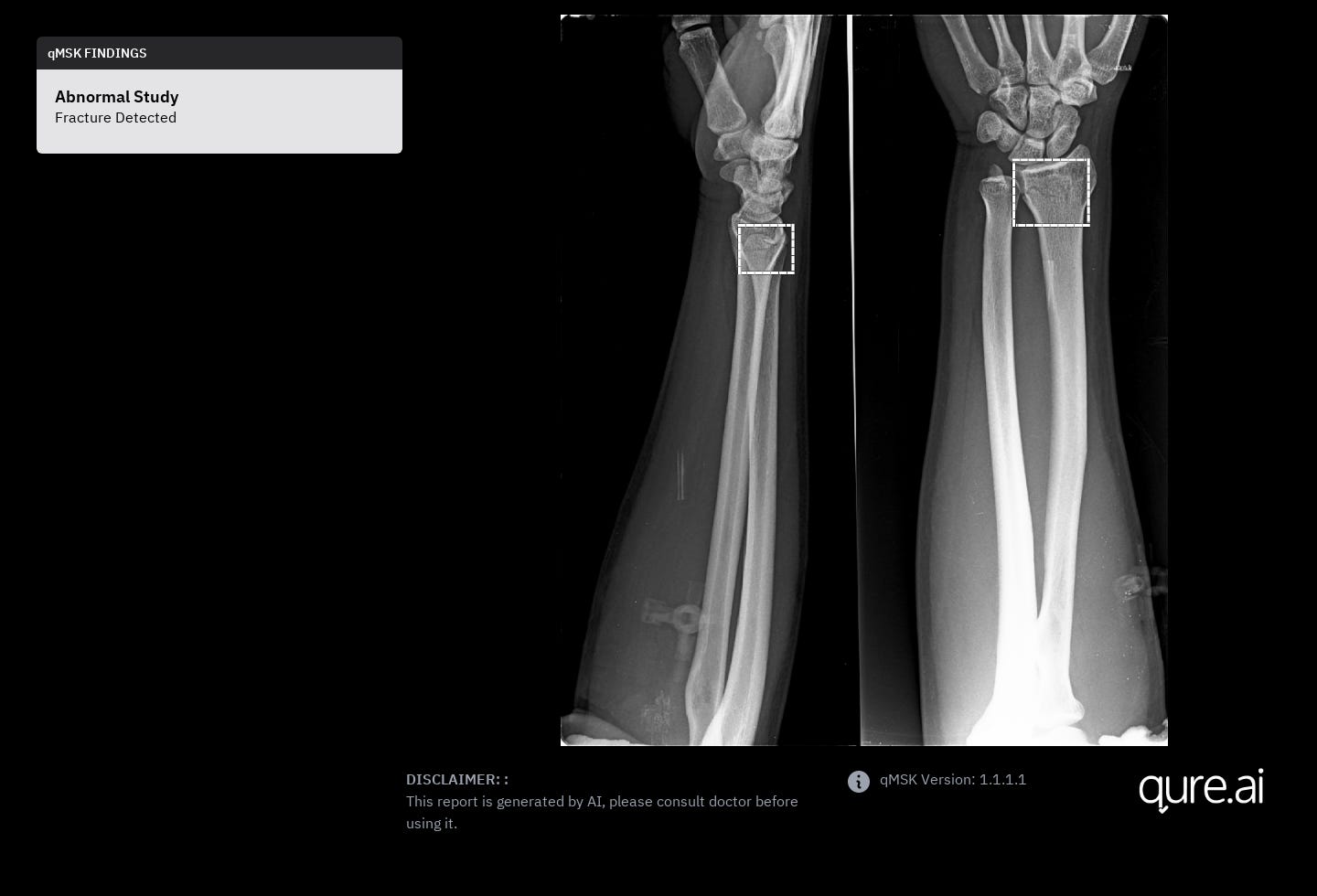
AI to Detect Fractures
Our recent study accepted for presentation in American College of Radiology (ACR) annual meeting highlights the potential of AI in improving the accuracy and efficiency of MSK imaging. In the study, we collected a massive dataset of 600,000 x-ray scans and their corresponding clinical radiology reports from various medical centers over the past year. The data was then annotated by radiologists and used to train a deep neural network, which was subsequently tested on a separate set of 150,000 x-rays.
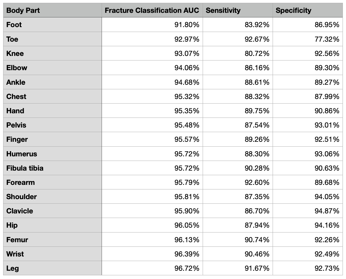
The AI model was trained for both classification and segmentation tasks, allowing it to accurately detect and isolate fractures in 16 different body parts. These fractures included various types such as healed, chronic, hairline, greenstick, acute, lateral, oblique, frontal, displaced, and undisplaced fractures. The deep neural network achieved remarkable results in detecting fractures in various body parts, achieving an overall AUC of 0.95 and a sensitivity and specificity of 0.8956 and 0.9273. The system demonstrated great performance in detecting fractures in the femur, leg, forearm, finger, hand, pelvis with AUC values greater than 0.93 and had lower performance for knee and foot.
It should be emphasized that the accuracy of AI algorithms for fracture detection is greatly influenced by the quality and quantity of the training data. Continuous efforts to collect and annotate large radiograph datasets are imperative to enhance the precision of these algorithms. Radiologists possess the expertise to identify and incorporate suitable AI software to assist non-radiologists in various situations. Moreover, they are also equipped to provide necessary oversight and training to other medical professionals regarding the use of AI tools for fracture detection.

AI is Radiology Extender
Furthermore, Dr. Amine Korchi, a radiologist from Geneva, highlights the importance of extending the use of AI beyond radiology to other healthcare specialties. In particular, emergency physicians could benefit greatly from AI-powered imaging technology, which could provide them with more accurate imaging information at the time of patient contact. This input, combined with the clinical examination and the physician's own judgment, can help render a more correct diagnosis and treatment approach the first time. This could also help triage patients and deliver initial care more safely and effectively, saving valuable time in the treatment pathway.

For AI to have a more widespread impact on the healthcare industry, it must be adopted beyond radiology. While most AI tools available today have been developed primarily for radiologists, they can be immensely useful when accessed by other specialists who may not have immediate access to a radiologist. It is important to note that AI support does not compete with radiologists as their tasks extend far beyond visual analysis of imaging studies. Instead, AI can augment their efficiency and allow them to focus on more complex, high-added-value tasks. By offering AI-based diagnostic support as a service, radiologists can increase their value to the healthcare enterprise and reduce the risk of incorrect clinical decisions.
Conclusion
In conclusion, AI has the potential to transform MSK imaging by providing diagnostic support to non-imaging specialists and augmenting the efficiency and value of radiologists. The development of accurate and reliable AI systems for MSK imaging will require ongoing research and development, as well as collaboration between radiologists and AI experts. With continued advancements in AI technology, we can expect to see significant improvements in the accuracy, efficiency, and accessibility of MSK imaging in the years to come.